
Pituitary adenoma may be demonstrated if involvement of the sella turcica is evident. The lambdoid suture is better evaluated than on nonangled views.

Mandible Radiographic Anatomy Wikiradiography Facial Bones Radiology Medical Anatomy
A-P at glabella â Protection.

. What is being demonstrated in the Townes skull. Ensure no rotation or tilt. How much is cr angle for both areas.
With forehead and nose resting on tabletop adjust head to place OML perpendicular to IR. Remove all metal plastic or other removable objects from the patients head. CR 30 deg caudad to OML or 37 deg caudad to.
Assessing the radiographs 40. PATIENT POSITIONRemove all metals plastics or other removable objects from the patients headPatient in erect or supinePART POSITION1. Learn vocabulary terms and more with flashcards games and other study tools.
Sunday February 23 2014. Occipital bone petrous pyramids and foramen magnum with dorsum sallae and posterior clinoids in its shadow are shown. Suture recognition on the AP view 44.
Moving or stationary grid. Immobilize the child with a bunny wrap. The central ray is angled ____ degrees _____ in the Townes AP axial of the skull.
Purpose and Structures Shown An additional view of the cervical spine. The addition of a Towne view to skull AP and lateral views has been thought to result in better sensitivity for detecting skull fractures than an AP and. The use of blocks and other radiolucent sponges will avoid exposing helpers.
The Townes SXR 39. Pediatric skull for craniosynostosis. The CR exits the _____ _____ in the Townes skull.
The AP frontal SXR 38. Depress chin and make sure OML is perpendicular to IR 2. PA 15 deg caldwell PA 25 deg or PA 0 deg.
Explain to the parents what you are going to do before you do it. 2-212 above the glabella. 70 to 80 kV.
No rotation or tilt midsagittal plane perpendicular to IR. For patients unable to flex their neck align. Basic positioning guidelines for AP townes view of the skull examining over and under angulation of the x-ray tube.
Elevate the shoulders using a firm pillow allowing the head to tilt backwards. Rest patients posterior skull against tableBucky surface. Make sure the child is naked from the waist up.
Is it a suture or a fracture. Supine without removing cervical collar if present. AP axial skull Towne method 2.
Start studying AP axial Townes Skull. Ap townes 30º caudad cr 25 above glabella both laterals both obliques. Depress chin and make sure OML is perpendicular to IR 2.
TOWNE METHOD skull series. Lie the child on the radiolucent sponge see below to place the IOML perpendicular the the IR. This is an alternative projection for patients who cannot flex their neck sufficiently for AP axial Towne.
Do image critique using PACEMAN guideline for the following projections. Seated erect or prone on table head aligned to CR and centerline of IR. Center IR to projected CR.
With possible spinal injury move patient to back edge of table and place IR about 1 25 cm below tabletop and posterior skull move floating tabletop forward. AP Axial Townes Skull CR Lines o _ Caudad OML _o Caudad IOML _ line perpendicular to IR _ Inches above glabella Right. The Towne view allows better frontal evaluation of the posterior fossa region than a standard nonangled frontal skull view.
Depressed chin OML perpendicular to IR. Use 14x17 and that will usually cover it. It results in magnification of the occipital area but results in lower doses to facial.
Take radiograph with patient in erect or supine position. Ap of skull down to the bottom of the shunt. Patient position and patient part.
Skull - Townes Trauma - also called Skull - AP Axial Area Covered. Align MSP to CR and to midline of the table3. View Skull positioningdocx from PA 0O at University of New England.
Ensure vertex of skull is within collimation field 4Ensure no rotation or tilt of headCENTERING. If not follow it all the way down. Fractures and pathologies of the skull.
Shield patients upper thoracic region. Ap townes 30º caudad cr 25 above glabella pa caldwell both laterals. PA axial haas SMV submentovertex where is the cr for an ap axial towne method.
Regular CR and DR as recommended by manufacturer. Align MSP to CR and to midline of the table3. PA axial facial Caldwell method Question.
24 x 30 cm Portrait. Center IR to horizontal beam CR to include entire skull. IR size 10 x 12 24 x 30 cm.
Suture recognition on the lateral view 42. CR is 25 in above glabella. PA axial facial Caldwell method Expert Solution.
Where does the central ray enter in the Townes AP axial of the skull. Ensure vertex of skull is within collimation field 4Ensure no rotation or tilt of headCENTERING POINTANGULATIONOFTUBEAngle 30 degrees caudad to OML orAngle 37 degrees caudad to IOMLtrauma caseCenter at MSP 25 inches65cm above the glabella. 8-9 PA axial15 Caldwell.
IR Size Orientation. AP axial skull Towne method 2. Film Screen Combination.
Suture recognition on the Townes view 41. Patient is erect or supine. Skull foramen magnum.
Up to 24 cash back Pediatric skull. 910 Basilar View of Sinuses â Measure.
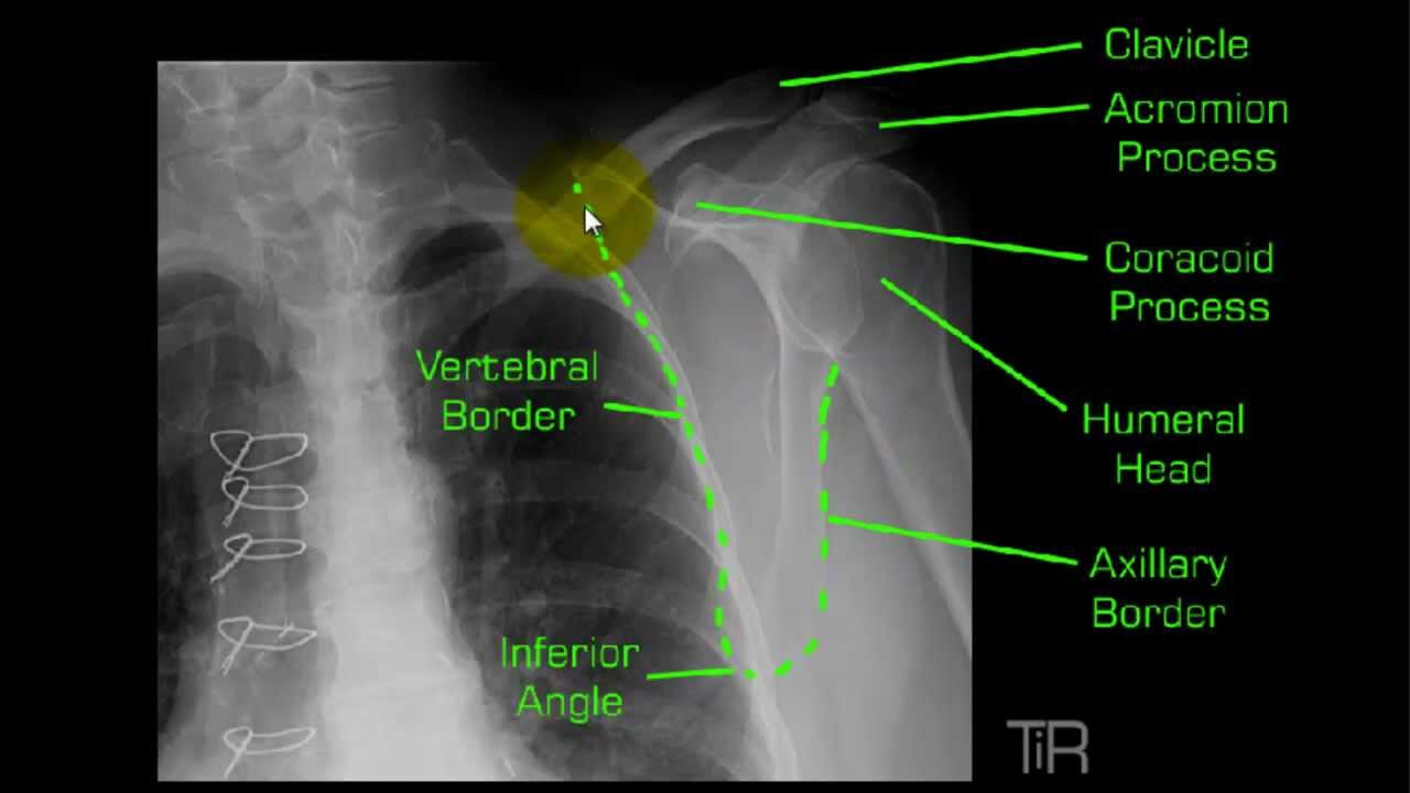
Y View Shoulder Mp4 Radiology Nursing Radiology Schools Anatomy


Mastoid Ap Axial Towne Methode Radiology Schools Radiology Cassette

Picture Skull Picture

Picture Skull Mirror Selfie Caldwell

Mountain Imaging Skull Positioning Radiology Schools Radiology Radiology Humor

Ap Skull Facial Bones Radiology Medical Radiography

Townes Skull Mp4 Radiology Technologist Book Worth Reading Medical

Cervical Spine Radiographic Anatomy Wikiradiography Diagnostic Imaging Radiology Radiology Schools

Punched Out Lesions In The Skull In A Case Of Multiple Myeloma Multiple Myeloma Myeloma Skull

Pin On Radiographs

Pin By Jamie L33 On Rad Tech Diagnostic Imaging Radiology Student Radiologic Technology
Pin On 服装店铺

Pin On Radiographs

Harlequin Eye Deformity Elevation Of The Superolateral Corner Of The Orbit It May Be Seen In Unilateral Plagiocephaly Or Bilate Sutures Harlequin Deformed

Standard Procedures For Skull Are Pa Caldwell Lateral Ap Axial Townes Radiology Imaging Radiology Student Radiography

Townes Skull Mp4 Radiology Technologist Book Worth Reading Medical

Pa Caldwell Skull You Know It S Pa Because Nostrils Are Defined There Isn T Equal Distance Between Orbital Margin And Skull M Radiographer V Shape Caldwell

Pin Em Cranio
